
Arterial cannulation for continuous blood pressure monitoring is not a technique free of complications, most of the time minor and not a major problem for the patient, but in some cases it can have serious consequences.
Furthermore, in children, given the smaller calibre of the arteries and the greater difficulty of cannulation, the incidence of these complications is greater, and for this reason there is currently a greater tendency to use ultrasound to perform the technique in an echoguided manner, reducing the incidence of these complications.
THE MAIN RISKS THAT MAY BE ENCOUNTERED DURING ARTERIAL CANNULATION ARE:
HEMATOMA
The risk of hematoma increases with the number of failed punctures. The use of ultrasound could help to reduce the incidence of hematoma.
INFECTIONS
The risk of infection increases in a time-dependent manner (10% if kept longer than 7 days). It is therefore recommended to change them periodically.
THROMBOSIS AND/OR OBSTRUCTION
Catheter obstruction in pediatric patients is very common due to their small caliber. Periodic flushing with 2-3 ml of saline solution can be performed, always in a gentle manner to reduce the risk of embolism. Thrombosis is especially common in umbilical artery catheters, so given the high risk of thrombosis and infection, we try to maintain them for a maximum of 48 hours.
We should remember that umbilical artery thrombosis can lead to necrotising enterocolitis, lower limb ischemia and renovascular thrombosis.
EMBOLISM
The embolism can become retrograde due to excessively vigorous and sustained lavage.
DISTAL ISCHEMIA
The severity of distal ischemia can vary from mild ischemia recoverable in a few days to complete ischemia requiring amputation, depending on the time of evolution and the distal flow preserved in the limb.
We can reduce the risk of ischemia by performing the Allen test prior to performing the technique if we are going to cannulate the radial artery to check the patency of the collateral circulation (ulnar). Furthermore, in pediatric patients it is important to select the correct caliber of catheter to be used, because if it is too large and does not allow sufficient blood to pass distally, the limb in question will develop ischemia if there is no collateral circulation.
INJURY TO ADJACENT NERVE STRUCTURES
The use of ultrasound will allow us to identify adjacent nerve structures, significantly reducing this risk.
IATROGENIC ARTERIOVENOUS FISTULA
An iatrogenic arteriovenous fistula occurs when there is a communication between artery and vein that results in continuous blood flow. Symptoms include constant pain at the puncture site, oedema, paresthesia, increased limb volume and tremor on palpation of the pulse.
POOR SIGNAL WAVEFORM AND DIFFICULTY IN IMPLEMENTING ADVANCED MONITORING
Another complication is the lack of an optimal blood pressure waveform signal and the difficulty in implementing advanced monitoring systems.
Apart from being a classic complication, the study of the arterial pressure waveform has become a complication of recent added value and very limiting.
RECOMMENDATIONS
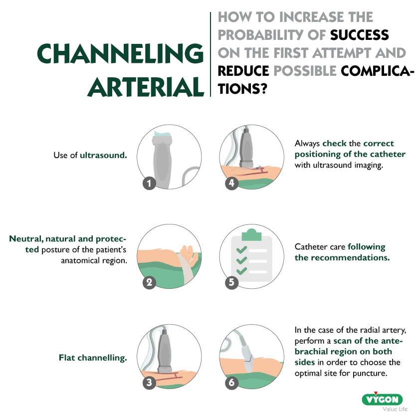
There are a number of actions that can be taken to increase the probability of success on the first attempt and thus reduce the potential risks:
- Use of ultrasound. This is the main recommendation, as it has been shown, both by meta-analysis and clinical practice, that the use of ultrasound for cannulation of peripheral arterial accesses decreases the complication rate.
- Neutral, natural and protected posture of the patient’s anatomical region.
- Flat channelling. Although it is technically more complex, it is always more reliable than cannulation out of plane, as the needle entry into the artery is better observed, as well as being able to observe if any complications arise, such as the appearance of a hematoma or dissection of the arterial wall when passing the guidewire.
- Always check the correct catheter placement by ultrasound imaging, blood gas sampling and blood pressure waveform recording.
- Catheter care following the recommendations specified in the Bacteremia Zero© protocol.
- In the case of the radial artery, perform a scan of the antebrachial region on both sides in order to choose the optimal site for puncture.
These recommendations, advices from Dr Escriba, anesthesist in Spain, will help to reduce the number of attempts during arterial cannulation and reduce the incidence of the complications described above.
Anesthesiologist at La Fe Hospital in Valencia, Spain.