
The reference guidelines on vascular access leave no doubt: it is essential to position and correctly locate the tip of a PICC. Otherwise, healthcare professionals risk encountering complications that can jeopardize the ongoing treatment and even the patient’s health.
To minimise these risks and accurately locate the tip of a PICC, four different methods can be used:
- X-ray
- Fluoroscopy
- Echocardiography (transthoracic or transoesophageal)
- ECG catheter tip location.
X-ray
It is an easily accessible resource in hospitals, either in the radiology department or at the bedside.
The catheter tip is located using anatomical references in the image.
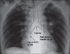
With this method, the cavoatrial junction cannot be seen and is therefore subjective.
This is shown in the study “Optimizing the patient positioning for PICC line tip determination” (Harako ME1, Nguyen TH, Cohen AJ), which proves that different professionals might interpret the catheter tip location in an X-ray quite differently.
The length of the Superior Vena Cava can vary considerably from one patient to another: between 4.4 and 10 cm, and this is only one of the anatomical structures which can change from one patient to another.
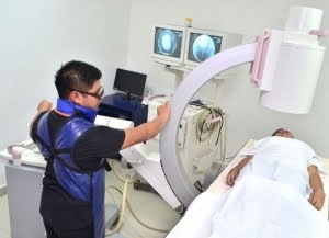
Fluoroscopy
This method, which involves X-rays, is mainly used in the interventional radiology department. Performing a real-time X-ray gives a continuous, live image of the patient. Catheter insertion can be visualised and the catheter tip can be located, to make sure it is in the right position.
Fluoroscopy is considered a sufficiently accurate method to position the central catheter. However, it is rather expensive, in terms of technology, equipment, training and logistics.
Its use is recommended only in special cases as it involves radiation exposure that should be avoided (INS).
Echocardiography
There are 2 types of echocardiography:
- Transesophageal (TEE)
- Transthoracic (TTE)
Transoesophageal (TEE) Echocardiography
Transesophageal echocardiography (TEE) can be very uncomfortable for the patient as it requires an upper endoscopy to place the probe close to the heart from inside the esophagus. Additionally, it necessitates the use of an operating room and trained personnel, limiting its application to very few patients.
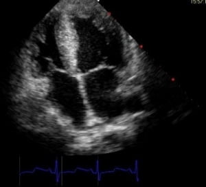
Transthoracic (TTE) Echocardiography
Compared to TEE, transthoracic echocardiography (TTE) is non-invasive and relies more on the technique used and the expertise of the professional. The ultrasound probe is placed on the patient’s chest at the level of the sternum to visualize the four cardiac chambers.
To accurately determine the position of the central catheter tip using echocardiography, a high level of knowledge in echocardiographic imaging is required.
Common points and differences between the two methods
There is no standardization of techniques; however, echocardiography is currently the most accurate method for locating the tip of a central line, surpassing the precision of fluoroscopy.
Regarding cost, an echocardiograph comes at a high price, though far from the investment required for a fluoroscope.
Both types of echocardiography are safe techniques but TEE is the more precise of the two.
ECG system
Although it is amortised in a relatively short period of time, this system involves a significant initial cost which is expensive compared to, for example, a single X-ray.
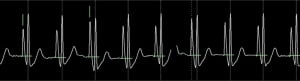
As with any ECG system, if it is located near other electrical devices, the image of the traces may contain artifacts.
Using this method, we place the electrodes on the patients body in the same way as for any standard ECG monitoring.
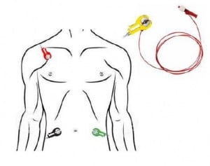
The intracavitary electrode (the red or yellow clip is connected to the catheter) allows the catheter tip to be accurately located thanks to the interpretation of the P-wave.
In fact, the most appropriate technique to locate the tip of a PICC is ECG. It is due to the following reasons:
- Easy to use: standard ECG monitor and minimal parameters
- Safe method: poses no additional risk compared to catheter placement itself
- Reliable and objective: observes changes in the P-wave, the technique is consistent for all
- Fast and cost-effective: an intraprocedural method (INS discourages subsequent procedures) and the system, in the long run, proves very affordable in terms of price.
Attempting to locate the tip of a PICC using an incorrect method can have negative consequences for both the professional and the patient. In the case of PICC, there is a demonstrated risk of:
- Thrombosis when it’s too short
- Arrhythmia when it’s too long
Bibliography
- Complicaciones del catéter según la posición de la punta Petersen et al, Am J Surg. 1999 Jul;178(1):38-41 Puel V et al, Cancer. 1993 Oct 1;72(7):2248-52 Eastridge et al, J Clin Oncol. 1995 Jan;13(1):233-8
- Cardiac Arrhythmias Resulting from a PICC: Two Cases and a Review of the Literature. Gapp J1, Krishnan M1, Ratnaraj F1, Schroell RP1, Moore D2. Cureus. 2017 Jun 3;9(6):e1308. doi: 10.7759/cureus.1308 ijccm.org/viewimage.asp?img=IJCCM_2010_14_4_180_76081_f6.jpg Aslamy et al, 1998? Sep;114(3):820-6 Chu et al, Anesth Analg.2004 Apr;98(4):910-4 Wirsing et al, Chest. 2008 Sep;134(3):527-33 Emerg Radiol.2004 Feb;10(4):186-9. Epub 2003 Dec 10
- Cardiac Arrhythmias Resulting from a PICC: Two Cases and a Review of the Literature. Gapp J1, Krishnan M1, Ratnaraj F1, Schroell RP1, Moore D2. Cureus. 2017 Jun 3;9(6):e1308. doi: 10.7759/cureus.1308.
- http://www.ijccm.org/viewimage.asp?img=IJCCM_2010_14_4_180_76081_f6.jpg
- Aslamy et al, 1998 Sep;114(3):820-6
- Chu et al, Anesth Analg.2004 Apr;98(4):910-4 Wirsing et al, Chest. 2008 Sep;134(3):527-33
- Emerg Radiol.2004 Feb;10(4):186-9. Epub 2003 Dec 10
- http://www.medyrad.net/rayos-x-con-fluoroscopia
- Journal of Infusion Nursing: infusion therapy standards of practice. Gebhard RE, Szmuk P, Pivalizza EG, Melnikov V, Vogt C, Warters RD. The accuracy of electrocardiogram-controlled central line placement. Anesth Analg 2007; 104: 65-70.
- Pittiruti M, Bertollo D, Briglia E et al. Intracavitary ECG method for positioning the tip of central venous catheters: results of an Italian multicenter study. J Vasc Access 2012; 13(3): 375-65.
- Pittiruti M, La Greca A, Scoppettuolo G. The electrocardiographic method for positioning the tip of central venous catheters. J Vasc Access 2011; 12(4): 280-291.
- Rossetti F, Pittiruti M, Lamperti M, Graziano U, Celentano D, Capozzoli G. Intracavitary ECG method for positioning the tip of central venous access devices in pediatric patients: results of an Italian multicenter study. J Vasc Access 2015; 16 (2): 137-143.