Neonatal patients admitted to Intensive Care Units are subjected to continuous manipulations and invasive procedures. One of the main objectives of nursing care is to minimize the practice of aggressive techniques in order to improve care and prevent complications.
However, despite the risks involved, there are some invasive procedures that are of vital importance in these units. Vascular access is undoubtedly one of them. Many patients who have undergone surgery or are critically ill require these devices for their survival. The use of intravascular therapies is increasingly employed in neonatal patients for multiple treatments.
Difficulties in central venous catheterization in neonates
Some newborns may present difficulties when it comes to cannulating the line, which makes certain “traditional” methods of insertion a real challenge in the care and protection of their venous capital. It is therefore essential for the nursing staff to look for alternatives to these techniques that allow the correct preservation of the vascular capital, which is indispensable for the future life of the neonatal patient.
Due to the small size of neonatal vessels, precise puncture and delicate insertion of the catheter are two critical steps in the method of vascular device placement. For this reason, we can affirm that central venous catheterization in neonates can be a demanding technique even for experienced physicians if they do not use an adequate cannulation technique.
For more than 20 years the method used in these patients has been known as the Seldinger Technique. Although this is a simple procedure well known to health care professionals, with high success rates in the adult population, in some neonatal patients this technique is not easy to apply and often requires several attempts before cannulation is achieved.
Given the small size of the vessels mentioned above, any error or mistake in the puncture can be fatal. The appearance of hematomas, for example, can hinder the possibilities of a second puncture attempt. In addition to this reality, mechanical complications such as obstruction or malposition of the catheter, closely related to the repeated number of attempts during the needle and guidewire insertion procedure, can also occur.
The risk of occurrence of mechanical complications increases with each additional attempt for cannulation of central devices in neonatal and pediatric patients. Therefore, succeeding on the first attempt is a priority in these small patients.
New methods for cannulating: the Seldinger Microseldinger Technique
Technological advances have allowed this technique to evolve. In 1989, Goodwin introduced the Modified Seldinger Method (MST) or Microseldinger Technique (micropuncture) in the insertion of peripherally inserted central venous catheters. Although this alternative technology is relatively new in the NICU – it has been used more extensively in adults – there are numerous studies that highlight its advantages over other methods in the high-risk neonatal population (preterm infants < 1000 grams or < 28 weeks gestation).
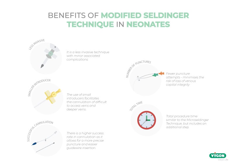
- Less invasive procedure with minor associated complications
- Allows the use of smaller introducers
- Higher cannulation success rate by allowing more precise puncture and easier guidewire insertion
- Fewer puncture attempts – minimizes risk of loss of venous capital integrity
- Total procedure time similar to the Microseldinger Technique, despite including an extra step.
The widespread use of this method has not been achieved due to the lack of devices and components suitable for the size of the patients admitted to the NICU, especially those with lower gestational age or weight. However, recent developments in the field of neonatology have made it possible to find systems suitable for performing MST even in the most vulnerable patients.
How to perform the Microseldinger or Modified Seldinger Technique?
The Microseldinger Technique consists of the use of an introducer that will serve as a guide to correctly route the catheter in the punctured vein. This introducer has a dilator inside it which, once removed, will allow the catheter to be inserted.
This insertion method has advantages in the placement of catheters both in neonates and in premature newborns of low birth weight. Its use is preferably indicated when one of the following circumstances is met:
- Blood vessels too small or in poor condition
- Veins difficult to visualize
- High risk of venous collapse
The use of this technique requires the introduction of an additional step to the Seldinger method. It is important that the professionals who are going to practice it have the appropriate education and training and know the protocols for performing micropuncture.
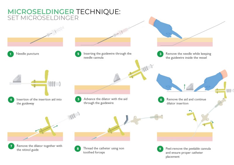
It starts with the puncture of the needle at an angle of 15-30º until blood return is observed. The nitinol guide is inserted and, while holding it, the needle is withdrawn. The dilator and cannula are advanced through the nitinol guide until it is inside the skin. The dilator and the nitinol guide are then removed and the cannula is inserted until it is inside the vessel. At this point, the catheter is inserted with the help of forceps until it reaches the appropriate position. Finally, the cannula is removed and the catheter is placed in the ideal position.
In the following infographic, to understand the technique correctly, you can see the step-by-step procedure:
Microseldinger technique and ultrasonography
The use of ultrasound technology in combination with MST has allowed, if possible, further improvements in terms of the ability to achieve venous access in complex patients and the reduction of pain during the cannulation process.
Echo-guided cannulation offers advantages to be able to observe and study a vessel, which, visually or by means of conventional cannulation, would not be possible. The use of ultrasound makes it possible to see how it is, in what condition, what size it is or what catheter can be applied to the vessel to be punctured.
This is fundamental, above all, when we are talking about:
- Patients in whom a central access is going to be placed as an emergency, i.e. patients weighing less than 1,500 grams or with pathologies that make them critical patients;
- Neonatal patients with prolonged accesses, who are expected to be admitted for a long time or who are going to undergo several surgeries and, therefore, need easier and safer accesses in order to preserve the venous capital.
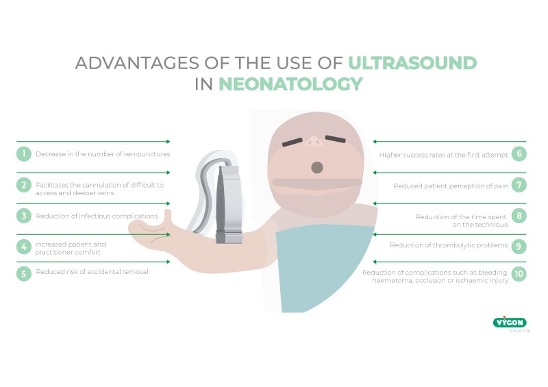
The use of ultrasound in combination with the micropuncture method facilitates cannulation of veins that are difficult to access, not visible or palpable, and veins of greater depth; it avoids endothelial trauma, thus reducing the possibility of thrombosis and further improving the cannulation success rate.
In addition, access to veins that are not normally visible has allowed healthcare professionals to use the middle third of the arm as the insertion point. The choice of this area brings with it a number of associated advantages:
- Reduction of infectious complications. The skin of the middle third of the arm is characterized by low bacterial colonization.
- Reduction of thrombotic complications, thanks to the possibility of measuring and using larger caliber veins.
- Greater comfort for the patient, due to the insertion site. Easier maintenance of the catheter and the exit site.
- Reduced risk of accidental removal because it can be better secured.
In conclusion, the studies currently available in neonatology on the use of the Microseldinger technique compared to the traditional one assure that the application of micropuncture is more beneficial in the neonatal population. Its combination with ultrasound technology, moreover, allows a higher success rate in cannulation and reduction of unnecessary multipunctures and associated consequences in vulnerable patients. For this reason, it is recommended that professionals be trained in the use of this technique in order to improve their care and facilitate better vital development.
Litterature
- A, H. Improving Neonatal Peripherally Inserted Central Catheter (PICC) Insertion: The use of Modified Seldinger Technique (MST). Retrieved from: https://galtmedical.com/wp-content/uploads/sites/10/2015/05/15GNT016_Microslide_White_Paper_SinglePages_HR_M.pdf
- Blanco Rodríguez, J., & Fernández Pérez, C. (2018). Ultrasound as a complementary method for the implantation of the peripherally inserted central venous catheter (PICC)(PhD). Complutense University of Madrid Faculty of nursing, physiotherapy and podiatry department of nursing.
- What are the latest recommendations on insertion by Seldinger technique, care and fixation of the peripherally inserted central venous line. Revised 6 May 2021, at https://www.murciasalud.es/preevid/19459.
- Mason Wyckoff, M., & Li Sharpe, E. (2015). Peripherally Inserted Central Catheters: Guidlne for Practice.(3rd ed., p. 50). Chicago: National Association of Neonatal Nurses. Retrieved from: http://hummingbirdmed.com/wp-content/uploads/sites/10/NANN15_PICC_Guidelines_FINAL.pdf
- Song, I., Kim, E., Lee, J., Jang, Y., Kim, H., & Kim, J. (2018). Seldinger vs modified Seldinger techniques for ultrasound-guided central venous catheterisation in neonates: a randomised controlled trial. British Journal Of Anaesthesia, 1332-1337(6). doi: 10.1016/j.bja.2018.08.008
Ajouter au projet Citavi par DOI.
- Tajuelo, I. (2020). Echo-guided cannulation in neonatal and preterm patients weighing less than 1,500 gr. . Reviewed at: https://campusvygon.com/global/canalizacion-ecoguiada-neonatos-prematuros/
In collaboration with
Sales representative for Vygon Spain in the Basque Country, Cantabria, Rioja, Navarra, Burgos and Soria.
EXPERIENCE
I have a degree in Communication Sciences, branch Journalism, but I have developed my entire professional career in the commercial sector, health and biotechnology.
I CAN HELP YOU IN…
I can offer you help in choosing the medical-surgical material you need for your health care work.
I am a specialist delegate in Intravascular Therapies and Oncology in Barcelona and Tarragona.
EXPERIENCE
Trained as a lawyer, I have been working for more than 20 years as a sales representative in a hospital environment providing service and support to health professionals so that they can offer the best service to the patient.
I CAN HELP YOU IN…
My work consists of listening, advising and training, both face-to-face and online, on VYGON products and the best techniques and procedures for their use.
A place to learn about health procedures and techniques from leading professionals.