

A Physiological Approach to Hemodynamic Management
Based on Pr. Pierre-Grégoire Guinot’s presentation at VYHEMDAYS 2025
Department of Anaesthesiology and Critical Care Medicine, Dijon University Medical Centre
Introduction
Each patient’s hemodynamic response is unique. In critically ill patients, failure to achieve proper tissue perfusion can determine outcome. The challenge lies in striking the right balance between fluid administration (which improves blood flow) and vasopressor use (which restores blood pressure): only the appropriate combination will ensure adequate perfusion without compromising organ function.
Current practice often relies on a “less fluids, more vasopressors” approach, driven by the fact that cardiac output is rarely monitored at the bedside. This leaves mean arterial pressure (MAP) as the primary treatment signal. However, raising MAP with vasopressors without identifying the underlying cause of hypotension may be harmful, as demonstrated by recent randomised trials: targeting 80-85 mmHg in older septic shock patients increased 90-day mortality and was stopped early for harm (Endo et al., 2025), and a meta-analysis of ten trials found no excess mortality with a permissive intraoperative target ≤ 60 mmHg (D’Amico et al., 2023). Conversely, fluid deficit can be as deleterious as fluid overload.
This article summarizes the key physiological principles needed to move from a reactive, pressure-targeted approach to a proactive, physiology-driven hemodynamic management strategy.
1. Understanding the Complexity of the Cardiovascular System
1.1. Beyond the “Simple Tube” Model
Traditionally, the cardiovascular system has been taught as a closed circuit where flow is governed by the pressure gradient between the arterial and venous compartments divided by systemic vascular resistance (SVR). This Ohm’s law analogy (MAP − CVP = CO × SVR) is an oversimplification with limitations. The cardiovascular system is composed of multiple interacting subsystems: the cardiac pump (right and left ventricles), the arterial tree (with elastic and resistive components), the microcirculation (the site of oxygen exchange), and the venous compartment (the capacitance reservoir). Cardiac output is not rigidly fixed but rather adapts to the metabolic oxygen demands of each tissue bed (Pinsky, 2002).
1.2. The Vascular Waterfall: A Paradigm Shift
A pivotal concept, often overlooked in standard teaching, is the vascular waterfall phenomenon. First described by Permutt and Riley (1963) in the pulmonary circulation, it has now been demonstrated in the systemic circulation at the bedside by Maas et al (2012).
▶ Key Concept: The Vascular Waterfall
When the critical closing pressure (Pcc) exceeds the mean systemic filling pressure (Pmsf), the arterial and venous circulations become functionally disconnected into two separate pressure systems. This is the vascular waterfall.
The critical closing pressure (Pcc) is the blood pressure below which flow ceases through small arterioles. It represents the vasomotor tone at the microcirculatory level. The Pcc partially reflect the vasomotor tone as it sums a tone-dependent wall tension term and the pressure surrounding the vessel. Usually, Pcc is approximately 25-50 mmHg and is significantly higher than Pmsf (~15–20 mmHg), creating a vascular waterfall.
The clinical implications are profound:
- Tissue perfusion pressure (TPP) = MAP − Pcc, not MAP − CVP. This means that the true driving pressure for organ perfusion depends on the relationship between MAP and the critical closing pressure, not on MAP alone. This gradient is macrovascular, and is not the same thing as microcirculatory perfusion (Sanchez et al., 2025).
- SVR calculated as (MAP − CVP)/CO becomes physiologically irrelevant in the presence of a waterfall, because it mixes two disconnected pressure systems (Maas et al., 2012).
- In vasoplegic states (e.g., sepsis, post-cardiopulmonary bypass), the waterfall nearly disappears. Norepinephrine can restore it in some patients improving cardiac index and tissue perfusion, yet it fails to do so in others, and no parameters can predict which patients would respond (Andrei et al., 2023).
- Capillary refill time tracks the arterial side of the waterfall: a baseline CRT > 3 s may be associated with altered vascular Waterfall, and ΔCRT correlated with changes of Pcc (Andrei et al., 2026).
2. Arterial hypotension Is Not the Disease: It Is always a Symptom of a Cause
Mean arterial pressure is the consequence of the interaction between cardiac output and vascular load. It always has an underlying cause, and treating hypotension with a vasopressor without identifying that cause can have serious consequences.
2.1. The Four Causes of Arterial Hypotension
When blood pressure is low, the underlying mechanism must be identified:
| Mechanism | HR | SV / CO | SVR | PPV/SVV | Treatment |
| Bradycardia | ↓↓ | SV → or ↑; CO ↓ | N or ↑ | Variable | Chronotrope (atropin) |
| Vasodilation | N or ↑ | N or ↑ | ↓↓ | N or ↑↑ | Vasopressor |
| Hypovolemia | N or ↑ | ↓ SV+CO | ↑ | ↑↑ | Fluids |
| Cardiac dysfunction (LV/RV failure) | N or ↑ | ↓↓ SV+CO | ↑↑ | Variable | Inotrope after confirmation |
It is essential to recognize that the cause of hypotension may change over time during surgery or critical illness. A patient who initially presents with vasodilation may subsequently develop hypovolemia or cardiac dysfunction. Continuous re-evaluation is mandatory and explain why patients should be monitored.
2.2. The U-Shaped Relationship Between MAP and Outcomes
Both too low and too high MAP are harmful. In 6,769 septic patients, a rising Pcc with a narrowing tissue perfusion pressure showed a U-shaped association with mortality and acute kidney injury (AKI), independent of MAP (Wang et al., 2026). The optimum is not universal: in elderly hypertensive surgical patients the lowest AKI rate was at 80–95 mmHg, not 65–79 mmHg (Wu et al., 2017). Attempting to achieve a higher MAP with increasing doses of vasopressors does not improve outcomes and may increase complications (Wu et al 2017).
Importantly, the dose of norepinephrine has been associated with occurrence of AKI (Guinot PG, Nguyen M, Evezard C, Fischer C, Bouhemad B. Dose-response association between intraoperative norepinephrine and postoperative acute kidney injury: a retrospective cohort study. Br J Anaesth. 2026 Jul 31:S0007-0912(26)00472-1. doi: 10.1016/j.bja.2026.07.001). This underscores that adequate blood pressure does not always guarantee optimal tissue perfusion or adequate cardiac output. Because norepinephrine raises MAP and Pcc together, a higher blood pressure maintained by vasopressor infusion does not by itself ensure a better perfusion: the gradient widens only if MAP rises more than Pcc (Bar et al., 2021).
3. The Pro-active Hemodynamic Optimization Strategy
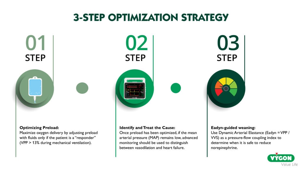
3.1. Step 1 — Optimize Preload First: Maximize Oxygen Delivery
The first fundamental step is to systematically optimize cardiac output by correcting preload. This increases oxygen delivery to tissues (DO₂ = CO × CaO₂).
Preload responsiveness should be assessed using dynamic indicators such as pulse pressure variation (PPV), stroke volume variation (SVV), or passive leg raising whenever PPV and SVV are not interpretable (spontaneous breathing, arrhythmia, low tidal volume, open chest). A PPV > 13% in mechanically ventilated patients (tidal volume ≥ 8 mL/kg, sinus rhythm; grey zone 9-13%) suggests that the patient is on the ascending limb of the Frank-Starling curve and may benefit from fluid administration.
3.2. Step 2 — Set the MAP Target, Then Identify and Treat the Cause of Hypotension
Once preload is optimized (i.e., the patient is no longer preload-responsive), if MAP remains below the objective target (e.g., 65–70 mmHg, or individualized for hypertensive patients), the next step is to identify why the blood pressure is low using the hemodynamic profile (Table 1). This is where advanced monitoring becomes essential: cardiac output differentiates vasodilation (normal CO, low SVR) from cardiac failure (low CO, high SVR), guiding the choice between vasopressor and inotrope.
3.3. Step 3 — Use Dynamic Arterial Elastance to Optimize Vasopressor Management
Dynamic Arterial Elastance (Eadyn) is defined as the ratio of pulse pressure variation (PPV) to stroke volume variation (SVV):
Eadyn = PPV / SVV
What does Eadyn reflect?
Eadyn is a functional index of the coupling between the pressure side and the flow side of the cardiovascular system. It reflects how efficiently the vascular system converts changes in stroke volume (flow) into changes in pulse pressure (pressure). In vasoplegic patients before treatment Eadyn is close to 0.9; norepinephrine lowers it to about 0.4 as it raises Pcc and arterial load (Bar et al., 2021). Crucially, Eadyn has been shown to correlate with the determinants of the vascular waterfall, specifically the critical closing pressure (Pcc), arterial compliance, and total peripheral resistance (Bar et al., 2021). This makes it an easy-to-read bedside index of arterial load, which moves with the tone-dependent component of Pcc without measuring it.
Clinical Application: Guiding Norepinephrine Weaning
▶ Key Point: Eadyn Is Used to Guide Vasopressor need during its use, Not for it’s initiation
Eadyn is NOT used to decide whether to start vasopressors. Its validated clinical role is to guide the reduction or weaning of norepinephrine in patients who are already receiving it.
The evidence is as follows:
- Eadyn above ≈0.9-0.95 (PPV > SVV): The vascular system maintains adequate pressure-flow coupling. The norepinephrine dose can be safely reduced step by step without anticipated significant MAP decrease (cut-off 0.94 in septic shock, Guinot et al., 2015; 0.90 by uncalibrated pulse contour, Bar et al., 2018).
- Eadyn below ≈0.9: The vascular system is unable to maintain pressure when flow changes occur. Reducing norepinephrine is likely to result in a clinically significant MAP decrease (>10-15%).
In the landmark SNEAD randomized trial (Guinot et al., 2017, Intensive Care Medicine), a hemodynamic algorithm based on Eadyn was associated with a shorter duration of norepinephrine treatment (17 h vs. 39 h, p < 0.001), a shorter ICU length of stay, a lower cumulative dose of norepinephrine, and no deterioration of perfusion parameters (lactate, ScvO₂). Later, a post-hoc analysis of the SNEAD study (Guinot et al., 2023) demonstrated that this Eadyn-guided norepinephrine weaning strategy was associated with a reduced incidence of acute kidney injury (27% vs. 51%, mainly lower stages).
Two caveats. Eadyn requires the same conditions as PPV and SVV, so it is uninterpretable in spontaneous breathing, arrhythmia or low tidal volume; and randomized trials applying it outside its validated indication have been negative (Guinot, 2026).
4. From Reactive to Proactive: A Paradigm Shift
Rather than reacting to hypotension, anticipate it. This requires:
- Continuous cardiac output monitoring to track blood flow before pressure drops
- Systematic assessment of preload responsiveness before administering fluids
- Identification of the mechanism of hypotension before treating it
- Use of Eadyn to guide safe vasopressor de-escalation and avoid unnecessary norepinephrine exposure
Conclusion
Achieving the right balance between flow (fluid) and pressure (vasopressors) requires a physiological, causal and individualized strategy, and means moving beyond MAP as the sole therapeutic target, towards the vascular waterfall, the determinants of venous return, and the coupling between pressure and flow.
Dynamic arterial elastance offers a practical, bedside-available tool to rationalize vasopressor management by predicting the hemodynamic response to norepinephrine dose changes. Its integration into clinical practice represents a significant step toward physiology-guided hemodynamic management, associated with reduced vasopressor exposure and improved renal outcomes.
The evolution from a passive, pressure-targeted approach to a proactive, physiology-driven response is not just a theoretical ideal—it is a clinical necessity.
Bibliography
Andrei, S., Bar, S., Nguyen, M., Bouhemad, B., & Guinot, P.G. (2023). Effect of norepinephrine on the vascular waterfall and tissue perfusion in vasoplegic hypotensive patients: a prospective, observational, applied physiology study in cardiac surgery. Intensive Care Medicine Experimental, 11, 52. https://doi.org/10.1186/s40635-023-00539-x
Andrei, S., Bar, S., Nguyen, M., Bouhemad, B., Longrois, D., & Guinot, P.G. (2026). Capillary refill time changes are associated with vascular waterfall response in post-cardiac surgery patients. Annals of Intensive Care, 16, 100105. https://doi.org/10.1016/j.aicoj.2026.100105
Bar, S., Nguyen, M., Abou-Arab, O., Dupont, H., Bouhemad, B., & Guinot, P.G. (2021). Dynamic Arterial Elastance Is Associated With the Vascular Waterfall in Patients Treated With Norepinephrine: An Observational Study. Frontiers in Physiology, 12, 583370. https://doi.org/10.3389/fphys.2021.583370
Bar, S., Leviel, F., Abou Arab, O., et al. (2018). Dynamic arterial elastance measured by uncalibrated pulse contour analysis predicts arterial-pressure response to a decrease in norepinephrine. British Journal of Anaesthesia, 121(3), 534–540. https://doi.org/10.1016/j.bja.2018.01.032
Chandrasekhar, A., Padrós-Valls, R., Pallarès-López, R., et al. (2023). Tissue perfusion pressure enables continuous hemodynamic evaluation and risk prediction in the intensive care unit. Nature Medicine, 29(8), 1998–2006. https://doi.org/10.1038/s41591-023-02474-6
D’Amico, F., Fominskiy, E.V., Turi, S., Pruna, A., Fresilli, S., Triulzi, M., Zangrillo, A., & Landoni, G. (2023). Intraoperative hypotension and postoperative outcomes: a meta-analysis of randomised trials. British Journal of Anaesthesia, 131(5), 823–831. https://doi.org/10.1016/j.bja.2023.08.026
Endo, A., Yamakawa, K., Tagami, T., et al. (2025). Efficacy of targeting high mean arterial pressure for older patients with septic shock (OPTPRESS): a multicentre, pragmatic, open-label, randomised controlled trial. Intensive Care Medicine, 51(5), 883–892. https://doi.org/10.1007/s00134-025-07910-4
Monge García, M.I., Gracia Romero, M., Gil Cano, A., et al. (2014). Dynamic arterial elastance as a predictor of arterial pressure response to fluid administration: a validation study. Critical Care, 18(6), 626. https://doi.org/10.1186/s13054-014-0626-6
Guinot, P.G. (2026). A plea for better understanding and appropriate use of dynamic arterial elastance: Lessons from negative results of randomised studies. European Journal of Anaesthesiology, 43(3), 199–201. https://doi.org/10.1097/EJA.0000000000002300
Guinot, P.G., Bernard, E., Levrard, M., Dupont, H., & Lorne, E. (2015). Dynamic arterial elastance predicts mean arterial pressure decrease associated with decreasing norepinephrine dosage in septic shock. Critical Care, 19, 14. https://doi.org/10.1186/s13054-014-0732-5
Guinot, P.G., Abou-Arab, O., Guilbart, M., et al. (2017). Monitoring dynamic arterial elastance as a means of decreasing the duration of norepinephrine treatment in vasoplegic syndrome following cardiac surgery: a prospective, randomized trial. Intensive Care Medicine, 43(5), 643–651. https://doi.org/10.1007/s00134-016-4666-z
Guinot, P.G., Huette, P., Bouhemad, B., Abou-Arab, O., & Nguyen, M. (2023). A norepinephrine weaning strategy using dynamic arterial elastance is associated with reduction of acute kidney injury in patients with vasoplegia after cardiac surgery: A post-hoc analysis of the randomized SNEAD study. Journal of Clinical Anesthesia, 88, 111124. https://doi.org/10.1016/j.jclinane.2023.111124
Maas, J.J., de Wilde, R.B., Aarts, L.P., Pinsky, M.R., & Jansen, J.R. (2012). Determination of vascular waterfall phenomenon by bedside measurement of mean systemic filling pressure and critical closing pressure in the intensive care unit. Anesthesia & Analgesia, 114(4), 803–810. https://doi.org/10.1213/ANE.0b013e318247fa44
Mulder, M.P. et al. (2023). Is Continuous Intraoperative Monitoring of Mean Arterial Pressure as Good as the Hypotension Prediction Index Algorithm? Anesthesiology, 138(6), 657–658. https://doi.org/10.1097/ALN.0000000000004541
Nguyen, M., Abou-Arab, O., Bar, S., et al. (2021). Echocardiographic measure of dynamic arterial elastance predict pressure response during norepinephrine weaning: an observational study. Scientific Reports, 11, 2853. https://doi.org/10.1038/s41598-021-82408-9
Pinsky, M.R. (2002). Functional Hemodynamic Monitoring: Applied Physiology at the Bedside. In: Vincent, J.L. (eds) Intensive Care Medicine. Springer. https://doi.org/10.1007/978-1-4757-5551-0_49
Sanchez, E.C., Taha, A., Tolba, Y., Hernandez, G., & Pinsky, M.R. (2025). Assessment of Tissue Perfusion Pressure in Patients With Septic Shock: Beyond Mean Arterial Pressure. Critical Care Medicine, 53(11), e2305–e2317. https://doi.org/10.1097/CCM.0000000000006805
Wang, J.Y., Diao, S.T., Zhu, T.Y., et al. (2026). Critical Closing and Tissue Perfusion Pressures in Sepsis: Implications for Risk Stratification — A Retrospective Cohort Study. Anesthesiology, 144(4), 886–897. https://doi.org/10.1097/ALN.0000000000005881
Wijnberge, M. et al. (2020). Effect of a Machine Learning–Derived Early Warning System for Intraoperative Hypotension vs Standard Care on Depth and Duration of Intraoperative Hypotension During Elective Noncardiac Surgery: The HYPE Randomized Clinical Trial. JAMA, 323(11), 1052–1060. https://doi.org/10.1001/jama.2020.0592
Wu, X. et al. (2017). Optimal blood pressure decreases acute kidney injury after gastrointestinal surgery in elderly hypertensive patients: A randomized study. Journal of Clinical Anesthesia, 43, 77–83. https://doi.org/10.1016/j.jclinane.2017.09.004