

Vascular access is the most frequent invasive procedure performed in hospitalized patients: more than 90% require intravenous therapy during their care management [1]. Globally, this accounts for over 2 billion peripheral intravenous catheter (PIVC) insertions every year [2].
However, despite being perceived as a routine procedure, it entails critical clinical challenges. Inadequate management or repeated punctures result in far more than temporary discomfort: they permanently impair the patient’s venous network, compromising future access and long-term care management [1].
In response, the international medical community is rethinking its practices around a structured, proactive approach: Vessel Health & Preservation (VHP) [14]. The objective goes beyond simply administering treatment; it involves adopting a more holistic vision of patient care, where sustainably protecting the integrity of the vascular system, whether involving peripheral or central access, is an intrinsic clinical priority.
I. Vascular Access: A Routine Procedure with Critical Consequences
The insertion of a peripheral intravenous catheter (PIVC) is a standard practice whose failure rates are well-documented in the literature: Helm et al. demonstrate that 35% to 50% of PIVCs fail before the completion of the prescribed therapy [3], while an international survey by Alexandrou et al. reveals that 14% of placed devices are ultimately never used for therapeutic purposes—precautionary insertions referred to in the literature as “idle catheters”—which unnecessarily expose patients to vascular trauma [2].
These failures are not without consequence. Each inappropriate or repeated insertion triggers a lesion cascade that extends beyond the puncture site. To understand why, one must examine the physical impact of each vascular breach on the vessel.
Vascular Homeostasis: A Finely Orchestrated Physiological Regulation
The vascular system is much more than a simple network of passive conduits; it is a fully functional organ. Its physiological equilibrium, known as vascular homeostasis, corresponds to the coordinated maintenance of several interdependent functions: vasomotor tone (the vessel’s ability to dilate or contract), wall permeability, blood fluidity, and the prevention of thrombosis. This balance is primarily orchestrated by the endothelium—the thin cellular layer lining the inner surface of all blood vessels—which actively regulates each of these mechanisms [4]. When preserved, the network fulfills its essential functions: nutrient transport, tissue oxygenation, and drainage. When compromised, all these functions become disorganized, disrupting the patient’s overall homeostasis.
What Each Puncture Triggers
Any breach of the vascular wall disrupts endothelial equilibrium and initiates a local inflammatory response. While physiologically normal in response to an isolated insult, this response becomes pathological when repeated. Epidemiological data attest to this: PIVC-related thrombophlebitis occurs in 25% to 35% of hospitalized patients carrying a catheter, leading to device removal, subsequent access difficulties, and prolonged hospital stays [5].
- On the venous side: Cumulative trauma leads to progressive and irreversible structural remodeling: fibrosis of the wall, intimal thickening, and luminal narrowing [1]. The peripheral network gradually becomes sclerotic, making each new puncture attempt more difficult than the last.
- On the arterial side: The challenges differ but remain significant: subjected to high pulsatile pressure, the arterial wall tolerates repeated trauma poorly.
Documented complications include thrombotic occlusion, hematoma, and, in the most severe cases, permanent ischemia of distal tissues that can progress to necrosis—complications for which identified risk factors include a high number of puncture attempts, pre-existing vascular pathologies, and the use of vasoconstrictors [6][7][8].
Consequences That Extend Beyond the Puncture Site
The repercussions of inadequate management do not stop at the punctured vessel. When local lesions progress without appropriate management, they can compromise prognosis on a much larger scale:
- Venous thrombosis, even when initially localized, exposes the patient to the risk of pulmonary embolism and a generalized pro-thrombotic state—a risk that the American College of Chest Physicians (ACCP) guidelines consider significant in the absence of treatment [9].
- Repeated or uncontrolled breaches can pave the way for bacteremia or even sepsis, which continues to carry a high mortality rate in hospital settings [5][10].
- Progressive depletion of the vascular capital compromises future access options, potentially delaying or preventing the administration of vital treatments in multimorbid, oncological, or chronic care patients [5][1].
Because of this systemic dimension, vascular access can no longer be viewed as a trivial procedure, but rather a dedicated clinical act that commits both prescribers and healthcare teams to a shared strategy of preservation.
II. Vessel Health and Preservation (VHP): A New Clinical Standard
This momentum has led to the development of a benchmark clinical framework: Vessel Health and Preservation (VHP). First formalized by Nancy Moureau and her collaborators in 2012 in the Journal of Vascular Access [11], it represents a new clinical standard in vascular access, founded on early assessment, reasoned device selection, qualified insertion, and continuous monitoring. It rests on a core principle: preserving the integrity of the vascular system is essential not only for immediate therapeutic efficacy but also for the viability of long-term access [1].
A Shift in Approach: Anticipating Rather Than Reacting
Where the traditional model allows device selection to be dictated by immediate clinical urgency or supply availability, this approach proposes a structured, anticipatory four-step process: early assessment of the vascular network, device planning, tailored insertion, and daily monitoring until removal [11]. Concretely, the model recommends planning within 24 hours of admission and insertion within 48 hours [1].
This individualized approach aligns clinical decision-making with each patient’s profile under the core principle: “the right device, in the right vessel, at the right time, taking into account the patient’s current and future needs” [21][22]. This strategy is also validated in specific settings. A single-center intensive care unit study on internal jugular central venous catheter insertion demonstrated that an organized, supervised approach improves first-attempt success rates and reduces the incidence of complications [12]. At the institutional level, deploying VHP also helps reduce material waste and strengthens infection control [1].
The Venous Network: A Non-Renewable Anatomical Capital
Each repeated or inappropriate breach leads to permanent structural remodeling—fibrosis, stenosis, thrombosis—that permanently reduces future access possibilities [1][11]. This capital is, by nature, finite and non-renewable: no healing process will restore a fibrosed or thrombosed vessel to its initial functional state. This is precisely why VHP places vascular preservation at the center of every access decision from the moment of patient admission.
Device Selection: A Patient-Therapy Equation
This decision relies on systematically matching two dimensions:
1. Patient characteristics: Vascular network quality and accessibility, puncture history, comorbidities, and risk profile.
2. Therapy requirements: Expected duration, physicochemical properties of the administered solutions (pH, osmolarity, vesicant nature), and diagnostic type.
This cross-analysis dictates the choice between a short peripheral catheter, long peripheral catheter, midline, or central access, consistently favoring the least invasive device appropriate for the situation [1].
This principle is now governed by benchmark international recommendations. The European ERPIUP 2023 consensus, developed by Pittiruti et al. under the auspices of the WoCoVA Foundation, proposes a classification of peripheral venous access devices according to their indication, duration of use, and compatibility with administered therapies, to standardize selection criteria internationally [13]. Similarly, the 2024 INS standards formalize early assessment and rigorous device selection as essential requirements to prevent inappropriate insertions, premature catheter changes, and vascular capital depletion [14].
By placing the preservation of the vascular network at the heart of clinical decision-making, VHP forms the clinical foundation for the practical objectives of the following sections: optimizing the first-attempt success rate and securing insertion via available technological and technical tools.
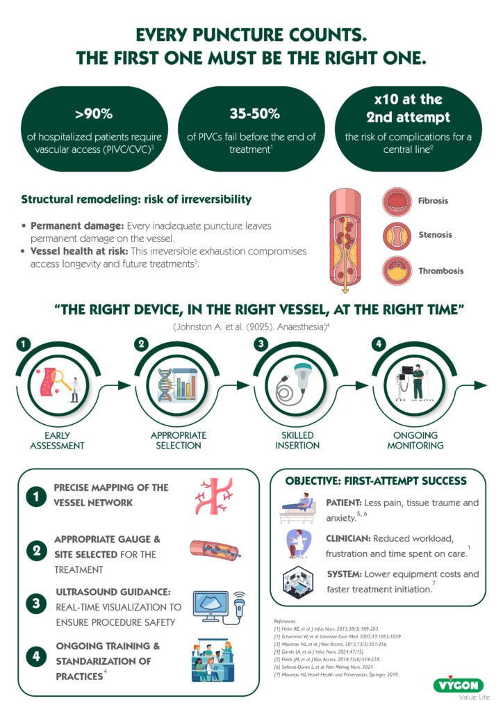
III. The First-Attempt Success Rate (FASR): The Clinical Benchmark Objective
In the field of vascular access, one indicator has progressively established itself as the central measure of procedural quality: the First-Attempt Success Rate (FASR). Both the 2024 INS standards [14] and the ERPIUP consensus [13] explicitly designate it as a priority objective, directly linked to vascular capital preservation and patient safety.
However, current clinical data reveal a significant gap between this objective and real-world clinical practice. Regardless of the hospital setting, a substantial proportion of initial PIVC insertion attempts fails, resulting in repeated and painful punctures. This ratio worsens further in DIVA (Difficult Intravenous Access) patients, where first-attempt failure rates range between 23% and 32% depending on the type of peripheral catheter used [16].
Each failure damages an additional venous site, restricts available options for subsequent attempts, and accelerates the exhaustion of an already compromised network. Consequences escalate with the number of attempts: according to a prospective study of 1,794 central venous catheterizations performed by experienced ICU operators, the risk of mechanical complications—hematoma, accidental arterial puncture, pneumothorax—increases 10-fold on the second attempt [17].
Objective Benefits at Three Levels
Adopting a proactive approach focused on the FASR yields documented benefits that extend beyond procedural comfort:
- For the patient: Reducing the number of attempts minimizes tissue trauma, the risk of hematoma and vascular spasm, as well as procedural pain and anxiety [18].
- For healthcare providers: Limiting repeated insertions reduces workload and material supply costs [19]. Furthermore, the mental load associated with repeated failures is a well-documented source of clinical frustration and burnout.
- For the healthcare system: Insertion failures delay necessary treatments and generate additional costs—an impact that quickly becomes substantial at an institutional scale [19].
Determinants of Success: What the Guidelines State
The FASR does not depend solely on technical dexterity; it is governed by upstream determinants that can be proactively managed. International guidelines, including the 2024 INS standards [14] and the ERPIUP consensus [13], identify four primary drivers:
- Rigorous prior assessment of the vascular network (both venous and arterial).
- Selection of the site and gauge suited to the therapy.
- Technical mastery of the procedure.
- The use of ultrasound guidance.
Additionally, a study conducted in the context of central catheterization identified risk factors for failure related simultaneously to the patient (obesity, history of chemotherapy, poorly visible or palpable veins), equipment, environment, and the method employed [12], underscoring the necessity of a systemic approach.
IV. Securing Insertion: The Alliance of Technology and Technique
When the vascular network is impaired or cannot be visualized, blind puncture exposes the patient to an increased risk of failure and additional trauma. Securing vascular access therefore relies on combining two complementary approaches.
Anatomical Exploration via Ultrasound
The 2025 guidelines of the American Society of Echocardiography recommend ultrasound guidance as a safety practice to improve cannulation success and minimize complications during all vascular access procedures—whether central, peripheral, or arterial—while noting that the quality of available scientific evidence remains limited to date [20].
In practice, ultrasound guidance allows clinicians to evaluate vessel depth and diameter, identify underlying anatomical variations, stenoses, thromboses, or calcifications beforehand, and optimize puncture site selection [20][21]. These benefits extend beyond immediate success: they help improve catheter lifespan while reducing infectious risks and procedural complications [20]. A recent meta-analysis of nurse-performed PIVC placements confirms the superiority of ultrasound guidance over the standard technique regarding success rates [23].
The Seldinger Technique
To secure catheter insertion following ultrasound-guided puncture, the Seldinger technique serves as a recognized mechanical standard. Introducing the device over a guidewire prevents the needle bevel from damaging the posterior vascular wall during advancement. This method preserves the vessel’s intima, limits parietal trauma, and ensures the longevity of the access route, particularly for long peripheral catheters, midlines, and arterial lines in intensive care settings [13][20].
Together, ultrasound guidance and the Seldinger technique form a powerful technical alliance serving a single goal: maximizing first-attempt success while safeguarding the patient’s vascular capital with every procedure.
V. Conclusion
The preservation of the vascular network is not a secondary issue—it is a transversal clinical requirement that involves all stakeholders in patient care. Prescribing physicians, care teams, and medical device manufacturers share a common responsibility: making every vascular access procedure a reasoned, planned, and minimally traumatic act.
The conceptual and technical tools are available. VHP provides the decision-making framework, ERPIUP and INS guidelines define objective selection criteria, and the alliance of ultrasound guidance with the Seldinger technique secures implementation. Their effective deployment relies on three prerequisites:
- Continuous training for healthcare teams,
- Institutional support at the facility level, and
- The integration of these practices into clinical service protocols.
Vascular access is not merely a procedure; it is a commitment to the patient’s healthcare trajectory. Every avoided puncture is a preserved access site. Every preserved access site is a maintained therapeutic option.
References
- Moureau N. (2025). Vessel health and preservation: Vascular access key risk considerations. Enfermería Clínica.
- Alexandrou E. et al. (2018). Use of Short Peripheral Intravenous Catheters: Characteristics, Management, and Outcomes Worldwide. Journal of Hospital Medicine.
- Helm R.E. et al. (2015). Accepted but unacceptable: peripheral IV catheter failure. Journal of Infusion Nursing.
- Félétou M. & Vanhoutte P.M. (2006). Endothelial dysfunction: a multifaceted disorder. American Journal of Physiology.
- Tagalakis V. et al. (2002). The epidemiology of peripheral vein infusion thrombophlebitis. The American Journal of Medicine.
- Brzezinski M. et al. (2009). Radial artery cannulation: a comprehensive review. Anesthesia & Analgesia.
- Kim S.Y. et al. (2016). Finger necrosis after accidental radial artery puncture. PMC.
- Ying Y. et al. (2022). Severe ischemia after radial artery catheterization: a literature review. Journal of Cardiovascular Anesthesia.
- Kearon C. et al. (2012). Antithrombotic therapy for VTE disease — ACCP Guidelines 9th ed. Chest.
- Mermel L.A. et al. (2009). Clinical practice guidelines for the diagnosis and management of intravascular catheter-related infection. Clinical Infectious Diseases.
- Moureau N.L. et al. (2012). Vessel Health and Preservation (Part 1): a new evidence-based approach to vascular access selection and management. Journal of Vascular Access.
- Liu Y. et al. (2022). Application of plan-do-check-act management to improve first-attempt insertion success rates of internal jugular vein catheterization for standardized training residents in an intensive care unit. PubMed.
- Pittiruti M. et al. (2023). European recommendations on the proper indication and use of peripheral venous access devices (ERPIUP). Journal of Vascular Access.
- Infusion Nurses Society. (2024). Infusion Therapy Standards of Practice. INS.
- Paterson R. et al. (2022). Peripheral intravenous catheter insertion in adult patients with difficult intravenous access: a systematic review. Emergency Medicine Australasia.
- Xu A. et al. (2024). Long guidewire peripheral intravenous catheters in emergency departments for management of difficult intravenous access. Academic Emergency Medicine.
- Schummer W., Schummer C., Rose N., et al. (2007). Mechanical complications and malpositions of central venous cannulations by experienced operators. A prospective study of 1794 catheterizations in critically ill patients. Intensive Care Medicine.
- Salleras-Duran L. et al. (2023). Pain and Satisfaction Perceptions of Ultrasound-Guided Versus Conventional Peripheral Intravenous Catheterization. Pain Management Nursing.
- Kleidon T.M. et al. (2025). A Prospective Cohort Study of Technique and Technology Used to Improve First Time PIVC Insertion Success. Journal of Advanced Nursing.
- Vegas A., Wells B., Braum P. et al. (2025). Guidelines for Performing Ultrasound-Guided Vascular Cannulation: Recommendations of the American Society of Echocardiography. Journal of the American Society of Echocardiography, 38(2), 57-91.
- Imbriaco G., Spencer T.R., Bardin-Spencer A. (2024). 10 best practice tips with radial arterial catheterization. Critical Care Medicine.
- Johnston A. et al. (2025). Association of Anaesthetists guidelines: safe vascular access 2025. Anaesthesia. doi: 10.1111/anae.16727.
- Tian Y. et al. (2024). The ultrasound-guided versus standard technique for peripheral intravenous catheter placement by nurses: a systematic review and meta-analysis. Heliyon, 10(9), e30582.