Fluid therapy is essential in the treatment of critically ill patients. Despite this, inadequate fluid management leads to significant morbidity and mortality.
WHAT WILL YOU FIND IN THIS ARTICLE?
- Fluid overload risks
- Cardiovascular overload
- Pulmonary oedema
- Mesenteric effects
- Myocardial Oedema
- Cutaneous Oedema
- CNS Effects
- How to avoid fluid overload?
- Individualised Approach
- Goal Directed Therapy
- Use of fluid response indicators
Want to know more about the risks associated with fluid overloading and how to avoid them? Stay on this page and read the full post.
Fluid therapy is essential in the treatment of critically ill patients. Despite this, inadequate fluid management leads to significant morbidity and mortality.
Selecting the right fluid and quantity for each of our patients is a challenge, there is no single, universal formula, it is an individualised treatment that requires a high degree of precision. However, there are guidelines that can help us to know the possible risks, avoid them and, if they occur, act early, improving the prognosis.
FLUID OVERLOAD RISKS
Cardiovascular overload and pulmonary oedema are the most common effects of fluid overload. However, there are other risks that are no less frequent but no less important, including mesenteric ischemia, cerebral oedema, coagulation defects, impaired tissue oxygenation and hypoproteinemia. 1
CARDIOVASCULAR OVERLOAD
One of the most common effects of excessive fluid administration is cardiovascular overload.1 In these situations, the heart is put under additional stress due to increased blood volume, which can lead to cardiac dysfunction and even heart failure, especially in patients with pre-existing heart disease.
PULMONARY EDEMA
The lungs are one of the organs where the adverse effects of fluid overload are most evident, which can lead to acute pulmonary edema or acute respiratory distress syndrome. 2
MESENTERIC EFFECTS
In situations of circulatory shock, the body redirects blood flow to vital organs such as the heart and brain. However, this leads to a decrease in blood flow to the digestive tract, which can result in mesenteric ischemia. Mesenteric ischemia leads to damage to the intestinal lining and causes loss of proteins and solutes into the intestinal lumen, which in turn contributes to loss of plasma volume. 2
MYOCARDIAL EDEMA
The massive administration of fluids in critical situations can lead to intravascular overload with the risks that this entails. When this occurs, the myocardium may favor edema formation, since, because of the increase in volume, both myocardial contractility and compliance are affected in the heart. 2
CUTANEOUS EDEMAS
Reduced plasma oncotic pressure can lead to systemic edema formation, which is clinically evident, especially after crystalloid resuscitation. Systemic edema not only has aesthetic implications but also results in decreased oxygen tension at that level and, consequently, can lead to ulcer formation and ulcer infection due to decreased cellular immunity. 2
CNS EFFECTS
Fluid administration can have significant effects on the central nervous system (CNS), particularly on the possibility of developing cerebral edema. The brain possesses protective mechanisms, such as the blood-brain barrier and vascular autoregulation, but in situations of shock, the critical decrease in plasma oncotic pressure can trigger an imbalance between hydrostatic and oncotic pressures in the brain bed, leading to cerebral edema. This underscores the importance of considering both the cause of brain damage and the integrity of the blood-brain barrier when administering large volumes of fluids. 2
HOW TO AVOID FLUID OVERLOAD?
Fluid therapy is a fundamental part of the treatment of the critically ill patient, however, as we have seen, in the event of fluid overload, the risks are serious.
Administering the exact amount that allows optimal treatment without exceeding the patient’s needs is a challenge, however, there are some recommendations that can help and guide us:
- Individualised Approach
- Goal Directed Therapy
- Use of fluid response indicators
How can each of these recommendations help us? In the following, we will address each of them.
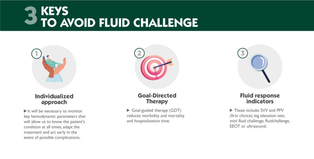
1. INDIVIDUALISED APPROACH
Every patient is different, and their needs will be different, so we must individualise therapy as much as possible. This will require monitoring of key hemodynamic parameters that will allow us to always know the patient’s condition, adapt the treatment and act early in the event of possible complications.
2. GOAL-DIRECTED THERAPY
Recent studies have shown that goal-directed therapy (GDT) can reduce morbidity and mortality and hospitalisation time. 3
This technique is based on continuous monitoring of hemodynamic parameters to guide treatment. In this way, it allows a more appropriate use of fluids, vasopressors and inotropes and therefore offers a better prognosis and faster recovery. 4
It has been shown that goal-directed perioperative therapy can improve postoperative outcome in high-risk surgical patients. 4
So much so that an unnecessary positive water balance is associated with increased morbidity and mortality. 5
Goal-directed therapy requires continuous monitoring of the patient’s hemodynamic changes. To do this, we will need an advanced hemodynamic monitoring system to inform us of the patient’s hemodynamic status accurately and reliably.
3. USE OF FLUID RESPONSE INDICATORS
Only 50% of critically ill patients respond to fluid administration by increasing their stroke volume (SV) and cardiac output (CO). To avoid life-threatening risks, it is essential to identify whether the patient is preload-dependent, i.e. responds to fluid infusion, or, on the contrary, fluid administration could lead to iatrogenesis, making the treatment not only futile but also potentially harmful.
Several techniques are available to determine whether the patient will respond to fluids, including:
- Systolic Volume Variation (SVV) and Pulse Pressure Variation (PPV). These are variables derived from the analysis of the arterial pressure waveform during mechanical ventilation and are considered a reliable predictor for measuring fluid responsiveness. Their high sensitivity for identifying fluid requirement and their strong relationship to cardiac index make these variables the first choice for assessing patient response to fluid resuscitation.
- Leg elevation test. This test involves transfusion of 300 ml of blood from the legs to the heart, changing the patient from a semi-reclined position to a 45° leg elevation. A good fluid response is considered if a >10% increase in systolic volume index is shown in the next minute.
- Mini fluid challenge test. Under continuous monitoring of cardiac output, administer a bolus of 100 ml of saline over one minute. If the cardiac output increases by more than 6%, the patient is considered to respond to fluid administration.
- Test fluid challenge. Similar to the previous one with the difference that the boluses administered are higher: 250ml in 10 minutes or 500ml in 20 minutes.
- End-expiratory occlusion test (EEOT). This consists of stopping mechanical ventilation at end-expiration for 15 to 30 seconds and measuring the resulting changes in cardiac output. As ventilation is stopped on expiration, the cyclic impedance in venous return is interrupted and cardiac preload increases. An increase in cardiac output above the 5% threshold is considered to indicate preload, and hence fluid responsiveness.
- Ultrasound. It is most commonly performed on the inferior vena cava, in which the diameter varies with changes in intravascular and intrathoracic pressure. That is, during inspiration the inferior vena cava collapses due to the negative pressure created by the expansion of the thorax. The patient will respond to fluids if diameter changes greater than 12% are shown.
Fluid overload is one of the risks we may encounter during fluid therapy in the critically ill patient, and as we have seen, complications can be very serious. Although challenging, there are techniques and tools that can help us avoid the potential risks.
A fundamental tool in these cases is hemodynamic monitoring. Having a hemodynamic monitor that provides us with constant, accurate and reliable target parameters will allow us to carry out goal-directed therapy and thus individualise treatment. In addition, it will also allow us to know if the patient is a fluid responder by using key variables such as Systolic Volume Variation (SVV) and Pulse Pressure Variation (PPV). This will improve the patient’s prognosis, allowing for more appropriate therapy and fewer complications, which, if they occur, will also be detected earlier.
BIBLIOGRAPHY
- Alfageme Michavilla, I., Alvarez, M. A., Alvarez Fernandez, J.A., Alvarez Marquez, E., Arias Garrido, J. J., Arnedillo Muñoz, A., Arroyo Maestre, J. M., Avellanas Chavala, m. L., ballesteros martínez, j. L., barranco medina, j., barranco ruiz, f., et al. (s. f.). Principios de Urgencias, Emergencias y Cuidados Críticos. UNINet. https://uninet.edu/tratado/c060208.html
- Claure-Del Granado, R., Mehta, R.L. Fluid overload in the ICU: evaluation and management. BMC Nephrol 17, 109 (2016). https://doi.org/10.1186/s12882-016-0323-6
- Chong, Matthew A.; Wang, Yongjun; Berbenetz, Nicolas M.; McConachie, Ian. Does goal-directed haemodynamic and fluid therapy improve peri-operative outcomes?: A systematic review and meta-analysis. European Journal of Anaesthesiology 35(7):p 469-483, July 2018. | DOI: 10.1097/EJA.0000000000000778
- Cannesson, M., Ramsingh, D., Rinehart, J., Demirjian, A., Vu, T., Vakharia, S., Imagawa, D., Yu, Z., Greenfield, S., & Kain, Z. (2015). Perioperative goal-directed therapy and postoperative outcomes in patients undergoing high-risk abdominal surgery: a historical-prospective, comparative effectiveness study. Critical care (London, England), 19(1), 261. https://doi.org/10.1186/s13054-015-0945-2
- Yang, X., & Du, B. (2014). Does pulse pressure variation predict fluid responsiveness in critically ill patients? A systematic review and meta-analysis. Critical care (London, England), 18(6), 650. https://doi.org/10.1186/s13054-014-0650-6
- Almela-Quilis, A., Millán Soria, J., Alonso Íñigo, J., & García Bermejo, P. (2015). Monitorización hemodinámica no invasiva o mínimamente invasiva en el paciente crítico en los servicios de urgencias y emergencias (27.a ed., Vol. 6). págs. 386-395. Emergencias: Revista de la Sociedad Española de Medicina de Urgencias y Emergencias. ISSN 1137-6821
- Marik, P.E., Monnet, X. & Teboul, JL. Hemodynamic parameters to guide fluid therapy. Ann. Intensive Care 1, 1 (2011). https://doi.org/10.1186/2110-5820-1-1
- Nava-López, J. A., & Bello-Melchor, M. (2013). Reanimación hídrica, parámetros hemodinámicos en anestesia. Revista Mexicana de Anestesiología, 36, 304-306. https://www.medigraphic.com/pdfs/rma/cma-2013/cmas131bt.pdf
A place to learn about health procedures and techniques from leading professionals.