
According to the studies by Dr Pittiruti and Dr LaGreca
Since the late 1980s, Dr Pittiruti and Dr LaGreca of the Fondazione Policlinico Universitario Agostino Gemelli in Rome have been researching the possibility of using ECG to locate the tip of a PICC.
In 1989 (1), they studied the feasibility of analysing the TQ segment to use this system in patients with atrial fibrillation.
In 2011, they succeeded in developing their own method to analyse the visible P waves on an intracavitary ECG for these cases (2).
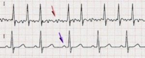
Interpretation of an ECG with AF
In an ECG trace, the P wave represents the synchronised depolarisation of the Right Atrium (RA) (blue arrow). An ECG system allows the position of the catheter tip to be determined by the interpretation of the P wave.
In the case of atrial fibrillation, there is no synchronised depolarisation (red arrow), so the TQ segment carries, not one, but several P waves (the sinoatrial node has anarchic electrical activity).
(3)
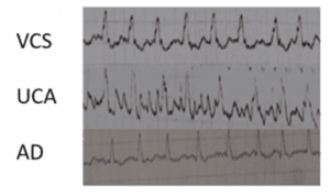
When there are several P waves but we want to place a central catheter using the ECG system, following the experts’ recommendations, we can use a modified ECG system and resort to the analysis of the TQ segment to determine the position of the catheter. This way, we can see the evolution of the P waves.
If we consider a heart with normal atrial activity, the cavoatrial junction is detected by a maximum P wave.
In the case of atrial fibrillation, when the catheter reaches the cavoatrial junction, the P waves of the TQ segment have maximum activity (frequency and height).
How to use modified ECG
(4) In 2016, Dr Pittiruti, Dr LaGreca and Dr Biasucci concluded that in the 10 cases studied, the method with modified ECG system is accurate (control with transoesophageal echocardiogram) through a thorough analysis which requires experience and time (5).
Shortly after the Steinhagen et.al study (6), they reached the same conclusions and suggested that the ECG method for patients with atrial fibrillation can eliminate the need for post-procedure verification.
Dr Pittiruti’s team wanted to establish clear parameters for interpretation of the TQ segment when the catheter is in the cavoatrial junction. They used three different parameters to measure the pattern of P waves in atrial fibrillation:
• The average of the P waves
• The height of the maximum P wave
• The difference between the maximum positive peak and the maximum negative peak of P waves
All three methods have proven to be effective in determining the exact position of the catheter.
The modified ECG system method has been shown to be safe in inserting and locating the central catheter tip in patients with atrial fibrillation.
However, analysis of the TQ segment is often difficult and takes time. For this reason, several years ago, an ECG system was developed to automatically perform such analysis.
In June 2019, Albertini et al. (7) published the first study on the use of the ECG system with atrial fibrillation mode (Pilot) and reached the following conclusions:
The ECG method, used as an intraprocedural control technique for positioning PICC type CVCs in patients with atrial fibrillation, is more reliable than anthropometric reference points. It is equally reliable whether the patient is diagnosed with atrial fibrillation or sinus rhythm.
At the 2018 WoCoVa congress, Dr LaGreca proposed this algorithm to determine which method is the most appropriate depending on the type of patient:

Bibliography
- Engelhardt W, Sold M, Helzel MV. ECG-controlled placement of central venous catheters in patients with atrial fibrillation. Anaesthesist 1989; 38: 476-9. https://pubmed.ncbi.nlm.nih.gov/2589630/
- Pittiruti M, La Greca A, Scoppettuolo G et al. EKG-controlled placement of central venous catheters in patients with atrial fibrillation (abstract). Oral presentation at the Annual Conference of the Infusion Nursing Society, May 21-26, 2011, Louisville (KY), USA.
- Heuser. Scheme of atrial fibrillation (top) and sinus rhythm (bottom). The purple arrow indicates a P wave, which is lost in atrial fibrillation
- Courtesy of Dr Pittiruti
- Pittiruti M, La Greca A, Biasucci DG et al. Intracavitary ECG for tip location in atrial fibrillation patients (abstract). Poster at the Annual Conference of the Association for Vascular Access, September 16-19, 2016, Orlando (FL), USA.
- Steinhagen F, Kanthak M, Kukuk G et al. Electrocardiography-controlled central venous catheter tip positioning in patients with atrial fibrillation J Vasc Access 2018, in press. https://pubmed.ncbi.nlm.nih.gov/29512399/
- Effectiveness of the ECG method in the correct positioning of PICC type central venous catheters in patients with atrial fibrillation Fabrizio Albertini*, Manuela Struglia, Vincenzo Faraone, Roberto Fioravanti, Stefano Boursier Niutta – Minerva Cardioangiologica 2019 June;67(3):207-13. https://www.minervamedica.it/en/journals/minerva-cardioangiologica/article.php?cod=R05Y2019N03A0207
A place to learn about health procedures and techniques from leading professionals.