

Central venous catheter (CVC) and peripherally inserted central catheter (PICC) placement are essential skills across acute and chronic care. Achieving an optimal tip position at the cavoatrial junction (CAJ) is critical for device performance and the prevention of complications, including arrhythmia, thrombosis and catheter dysfunction. Traditionally, tip confirmation has relied on post-procedural chest radiography (CXR). However, over the past two decades, intracavitary electrocardiogram (IC-ECG) guidance has emerged as a reliable, real-time alternative that is increasingly supported by evidence and clinical practice.
This article explores the principles of ECG-guided tip positioning, the supporting evidence base, and its role in contemporary vascular access pathways.
The principle of ECG-guided tip positioning
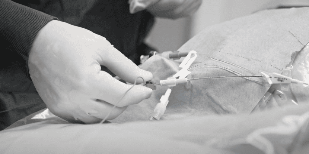
ECG-guided catheter placement uses the patient’s intrinsic cardiac electrical activity to identify the catheter tip location in real time. The technique typically involves:
- Connecting the catheter (or guidewire/saline column) to an ECG monitor
- Observing changes in the P wave amplitude as the tip approaches the CAJ
- Identifying the point of maximal P wave amplitude, which correlates with optimal tip position
As the catheter advances into the superior vena cava (SVC) and towards the right atrium, the P wave progressively increases in amplitude. Entry into the right atrium is typically associated with a biphasic or negative deflection, signalling the need to withdraw slightly.
This physiological feedback allows clinicians to position the catheter accurately during insertion, rather than relying on estimation followed by radiographic confirmation.
Evidence supporting ECG guidance
Early work by Pittiruti et al. (2008) [1] and Moureau et al. (2010) [2] demonstrated that ECG-guided PICC placement is highly accurate and can reduce or eliminate the need for routine CXR confirmation when appropriate protocols are followed. These studies showed strong correlation between maximal P wave amplitude and correct CAJ positioning, supporting the reliability of the method.
Subsequent studies continue to reinforce these findings:
- ECG-guided techniques have been shown to significantly reduce malposition rates compared with traditional landmark-based insertion. In one study, no malpositions were observed in the ECG group compared with 18% using anatomical landmarks. [3]
- Arrhythmias and over-insertion events were also reduced when ECG guidance was used, highlighting improved procedural safety. [3]
More recent data demonstrates improved accuracy when ECG is combined with ultrasound guidance, achieving correct tip placement rates exceeding 90%. [4]
- A 2024 comparative study found that modern ECG-guided techniques achieve clinically acceptable tip positioning with minimal variation between methods. [5]
- A 2025 meta-analysis in neonatal care confirmed that intracavitary ECG improves tip accuracy while influencing complication profiles depending on technique used. [6]
Reducing reliance on chest radiography
The move away from routine CXR is a key driver for ECG adoption:
- Radiographic confirmation remains widely used, but there is growing evidence supporting ECG- and ultrasound-based protocols as safe alternatives. [4]
- ECG-guided placement enables immediate confirmation at the bedside, reducing delays in catheter use and streamlining workflow
- It also avoids radiation exposure and reduces resource utilisation
Emerging innovations, including artificial intelligence-assisted ECG interpretation, are being explored to further enhance accuracy and reduce operator variability. [7]
Clinical advantages of ECG-guided placement
1. Real-time positioning
Immediate feedback during insertion allows precise tip placement without delay.
2. Improved patient safety
Lower rates of malposition, arrhythmia and catheter-related complications have been reported.
3. Workflow efficiency
Eliminates the need to wait for radiographic confirmation in many patients, enabling faster therapy initiation.
4. Reduced radiation exposure
Avoids unnecessary imaging, particularly beneficial in populations requiring repeated access.
5. Cost-effectiveness
Reduced imaging and fewer repositioning procedures can lower overall costs.
Considerations and limitations
While ECG guidance is widely applicable, it is not suitable for all patients. Limitations include:
- Atrial fibrillation or absent/indistinct P waves
- Pacemaker dependency
- Certain cardiac conduction abnormalities
In such cases, alternative confirmation methods such as imaging remain necessary.
Operator training is also essential, as interpretation of ECG waveforms and integration into clinical workflow requires competency and standardisation.
Integrating ECG guidance into practice
Best practice increasingly involves a multimodal approach:
- Ultrasound for vessel selection and cannulation
- ECG guidance for real-time tip positioning
- Selective imaging when indicated
This integrated pathway aligns with evolving vascular access recommendations and supports safer, more efficient care.
Devices and systems designed to facilitate ECG-guided placement, including dedicated tip confirmation technologies such as Combcard, can support clinicians by simplifying set-up and signal acquisition. When used within a structured protocol, these systems may enhance consistency and ease of adoption.
Future directions
The field continues to evolve, with current research focusing on:
- AI-enhanced ECG signal interpretation
- Integration with digital vascular access platforms
- Standardisation of training and competency frameworks
- Expanded use in complex patient populations
As evidence grows, ECG-guided placement is likely to become a routine component of bedside vascular access, particularly for PICCs.
Conclusion
ECG-guided central venous catheter placement represents a significant advancement in vascular access practice. By enabling accurate, real-time tip positioning, it reduces reliance on radiographic confirmation, improves patient safety and enhances procedural efficiency.
Supported by a robust and growing evidence base, and complemented by technologies that facilitate its use, ECG guidance is well positioned as a cornerstone of modern catheter placement pathways.
ECG-Guided Central Venous Catheter (CVC) Tip Placement: A Safer, Faster, and Cost-Effective Alternative
Read to discover the ECG-Guided Technique: A Step-by-Step Guide
References
- Pittiruti M, et al. The ECG method for positioning the tip of PICCs: results from two preliminary studies. JAVA. 2008;13(4):112–119.
- Moureau N, et al. Electrocardiogram (EKG) Guided Peripherally Inserted Central Catheter Placement and Tip Position: Results of a Trial to Replace Radiological Confirmation. JAVA. 2010;15(1):9–15.
- Krishnan AK, et al. Electrocardiogram-guided technique: an alternative method for confirming central venous catheter tip placement. J Emerg Trauma Shock. 2018;11(4):276–281. [ncbi.nlm.nih.gov]
- Jiang X, et al. Safety and feasibility of ECG-guided tip positioning combined with ultrasound-guided PICC placement. 2024. [link.springer.com]
- Gullo G, et al. Comparison of ECG guidance techniques for PICC insertion. Sensors. 2024. [ivteam.com]
- Wang Z, et al. Intracavitary ECG guidance for PICC placement in newborns: a network meta-analysis. Medicine (Baltimore). 2025. [pmc.ncbi.nlm.nih.gov]
- Lee K, et al. Evaluation of intravenous ECG for PICC placement and AI-enhanced accuracy. Br J Anaesth. 2025. [bjanaesthesia.org]
- Rethinking routine chest radiography after central venous catheter placement. 2026. [link.springer.com]

A place to learn about health procedures and techniques from leading professionals.