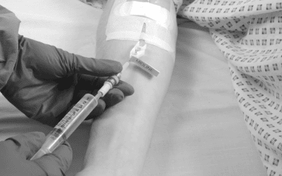
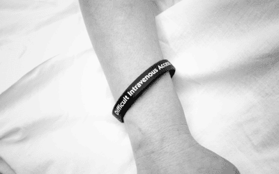
Ultrasound-guided vascular access has become a cornerstone of modern clinical practice, offering a safer, more accurate alternative to traditional landmark-based techniques. First described in 19781, the use of ultrasound for central venous catheterisation has evolved into a widely accepted standard, particularly for patients with difficult intravenous access (DIVA).
Why Ultrasound Matters in Vascular Access
Ultrasound imaging allows clinicians to visualise veins and surrounding structures in real time. This capability significantly improves the accuracy of catheter placement and reduces the risk of complications such as arterial puncture, nerve damage, or haematoma. It also enhances first-pass success rates and shortens procedure times.
Key benefits of ultrasound-guided vascular access include:
- Improved safety: Fewer mechanical complications and reduced risk of misplacement.
- Higher success rates: Increased likelihood of successful placements on the first attempt.
- Time efficiency: Shorter procedure times and fewer repeated attempts.
- Cost-effectiveness: Reduced need for additional interventions and associated resources.
Applications Across Vascular Access Sites
- Internal Jugular Vein: Ultrasound guidance reduces complications and improves success rates compared to landmark techniques.
- Subclavian Vein: Studies show that real-time ultrasound significantly outperforms traditional methods in terms of safety and efficiency.
- Femoral Vein: Ultrasound use decreases the number of needle passes and minimises complications such as arterial puncture.
Midline Catheter Placement
Ultrasound is particularly valuable for midline catheter insertion, especially in patients with DIVA. Recently, insertion of a midline catheter is recommended to be performed under ultrasound guidance and placed in the arm2. A study of DIVA patients in the surgical intensive care unit has shown that ultrasound-guided midline catheters are not only cost-effective, but can help facilitate early central line removal and, therefore, may reduce central line-associated bloodstream infections (CLABSI) rates3.
With proven cost savings for the NHS4, midlines are becoming increasingly popular. Using ultrasound to assist in midline placement could bring additional benefits to hospitals and Trusts.
References:
[1] Ullman JI, Stoelting RK. Internal jugular vein location with the ultrasound Doppler blood flow detector. Anesth Analg. 1978 Jan-Feb;57(1):118. doi: 10.1213/00000539-197801000-00024. PMID: 564628.
[2] Yokota T, Tokumine J, Lefor AK, Hasegawa A, Yorozu T, Asao T. Ultrasound-guided placement of a midline catheter in a patient with extensive postburn contractures: A Case report. Medicine (Baltimore). 2019 Jan;98(3):e14208. doi: 10.1097/MD.0000000000014208. PMID: 30653177; PMCID: PMC6370112.
[3] Deutsch GB, Sathyanarayana SA, Singh N, Nicastro J. Ultrasound-guided placement of midline catheters in the surgical intensive care unit: a cost-effective proposal for timely central line removal. J Surg Res. 2014 Sep;191(1):1-5. doi: 10.1016/j.jss.2013.03.047. Epub 2013 Apr 6. PMID: 24565504.
[4] Pioneering Midline Catheter from Vygon Saves NHS Trust Over £75k. 2023 May 9th. http://vygon.co.uk/midline-catheter-gloucestershire-case-study/