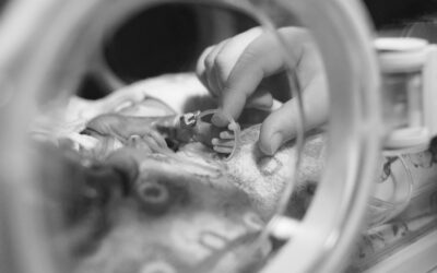

Umbilical catheterisation remains a cornerstone of neonatal intensive care, providing reliable vascular access for newborn infants who require medication, fluids, parenteral nutrition, blood products, or close haemodynamic monitoring. When performed by trained clinicians using a structured and evidence-based approach, both umbilical venous catheters (UVCs) and umbilical arterial catheters (UACs) can facilitate timely treatment while supporting patient safety.¹,²
Although catheter insertion is a common procedure in neonatal practice, success relies on more than technical skill alone. Careful preparation, accurate tip positioning, secure fixation, and ongoing surveillance all contribute to reducing the risk of complications and promoting optimal outcomes.¹,³
The Role of Umbilical Catheters in Neonatal Care
Umbilical vascular access is most commonly established during the first days of life and is particularly valuable in preterm and critically ill infants. UVCs provide central venous access for the administration of medications, concentrated glucose solutions, parenteral nutrition, inotropes, and emergency therapies, while UACs support continuous blood pressure monitoring and frequent blood sampling.¹,²
The ability to establish dependable vascular access quickly can support clinical decision-making and reduce the need for repeated peripheral cannulation, helping to minimise stress for both infants and care teams.²
Preparation: Setting Up for Success
Successful catheter placement begins before the procedure starts. Clinicians should confirm the indication for insertion, review any contraindications, and ensure appropriate equipment is available. Relative contraindications may include abdominal wall abnormalities, omphalitis, necrotising enterocolitis, or significant local infection.³,⁴
Aseptic technique remains fundamental to reducing the risk of catheter-related infection. Appropriate hand hygiene, sterile barriers, skin preparation according to local policy, and maintenance of a sterile field are all important considerations. Attention should also be paid to thermoregulation, monitoring, infant positioning, and comfort throughout the procedure.¹,²
Procedural Overview
While insertion techniques vary slightly between organisations and should always follow local guidelines, several key principles are widely recognised.
1. Preparation
- Confirm indication and suitability for catheterisation.
- Estimate insertion depth before beginning the procedure.
- Assemble equipment and establish monitoring.
- Prepare and drape the field using aseptic technique.¹,³
2. Vessel Identification
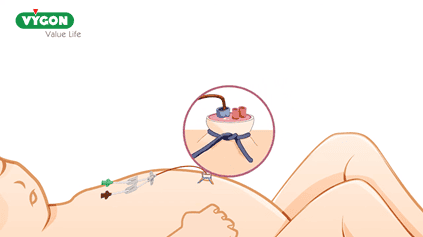
- Identify the target vessel within the umbilical stump.
- Ensure adequate visualisation before attempting cannulation.
- Maintain gentle tissue handling throughout the procedure.⁵,⁶
3. Catheter Advancement
- Advance the catheter carefully to the anticipated depth.
- Avoid forcing the catheter if resistance is encountered.
- Reassess vessel position and pathway if advancement proves difficult.⁵,⁶
4. Position Confirmation
- Confirm catheter tip position using imaging in accordance with local policy.
- Consider ultrasound where available to support accurate visualisation.
- Verify appropriate tip location before commencing intended therapies.²,⁶
5. Securement and Documentation
- Secure the catheter using approved fixation methods.
- Record insertion depth, tip position, and any procedural observations.
- Ensure clear communication during clinical handover.¹,³
6. Ongoing Monitoring
- Evaluate line function and necessity daily.
- Monitor for migration, infection, thrombosis, extravasation, or vascular compromise.
- Remove the catheter when central access is no longer clinically required.¹,²,³
Achieving Optimal Tip Position
Correct catheter tip placement is one of the most important determinants of safety.
For UVCs, the catheter should pass from the umbilical vein through the ductus venosus and terminate within the inferior vena cava at the level of the diaphragm. Guidance commonly recommends a tip position around the T8-T9 vertebral level and outside the cardiac silhouette. Placement that is too low or within the portal venous system may increase the risk of complications, while intracardiac positioning should be avoided.²,³,⁶
For UACs, accepted tip positions are typically either a high thoracic position or a low abdominal position, with many guidelines favouring high placement because of a lower risk of vascular complications.⁷,⁶
Clinical Pearls for Successful Umbilical Catheter Placement
Plan before you insert
Calculate the anticipated insertion depth before beginning and ensure equipment is prepared and organised. Good preparation can help reduce procedural time and minimise unnecessary manipulations.¹,³
Maintain a strong focus on asepsis
Catheter-related infection remains one of the most significant preventable complications associated with umbilical lines.²,³
Never force advancement
Resistance may indicate entry into a side branch or an incorrect pathway. Reassessment is generally preferable to applying additional force.⁵,⁶
Aim for the ideal tip position from the outset
Accurate placement can reduce the need for repositioning and may lower the risk of complications associated with malpositioned catheters.²,³
Confirm position before use
Radiographic and, where available, ultrasound confirmation can provide reassurance that therapies are being delivered through an appropriately positioned catheter.²,⁶
Remember that catheters can migrate
A line that is correctly positioned immediately after insertion may move over time, making ongoing assessment important.²,³
Be alert to subtle warning signs
Abdominal distension, altered perfusion, oedema, unexpected clinical deterioration, or difficulties with aspiration should prompt further assessment.³,⁷
Review daily and remove promptly
Daily evaluation of line necessity supports good vascular access stewardship and may help reduce infection risk. 1,2,3
Supporting Best Practice
Umbilical catheter placement is a valuable skill that continues to play an important role in neonatal care. A combination of anatomical knowledge, careful preparation, meticulous aseptic technique, accurate tip placement, and ongoing monitoring can help clinicians maximise the benefits of vascular access while minimising risk. By following local guidance and maintaining procedural competency, neonatal teams can continue to support safe and effective care for vulnerable newborn infants.1,2,5
References
- Royal Cornwall Hospitals NHS Trust. Umbilical Line Insertion (UAC, UVC) Neonatal Clinical Guideline. Version 5.0. March 2025. [doclibrary…all.nhs.uk]
- East of England Neonatal Operational Delivery Network. UVC Guideline. June 2025. [eoeneonata…ork.nhs.uk]
- Ashford and St Peter’s Hospitals NHS Foundation Trust. Umbilical Venous Catheterisation Guideline. January 2025. [ashfordstpeters.net]
- NHS Lanarkshire. Umbilical Venous Catheter Guideline. Version 12. 2020. [rightdecis…cot.nhs.uk]
- NHS Greater Glasgow and Clyde. Umbilical Catheters Guideline. Neonatology Clinical Guidelines. [clinicalgu…cot.nhs.uk]
- van Schuppen J, Onland W, van Rijn RR. Lines and Tubes in Neonates. The Radiology Assistant. 2013. [radiologya…sistant.nl]
- Ashford and St Peter’s Hospitals NHS Foundation Trust. Umbilical Artery Catheter Insertion Guideline. February 2025. [ashfordstpeters.net]

A place to learn about health procedures and techniques from leading professionals.